The human foot supports your entire body and enables mobility. Each step you take requires a complex system of bones, muscles, tendons, and ligaments to work together. By understanding foot anatomy, you can better care for your feet, prevent injuries, and seek help when something feels off. At ModPod Podiatry, we focus on keeping your feet healthy, helping you move comfortably, and treating issues that may arise.
This guide will break down the anatomy of the foot, explain its functions, and show how foot problems can develop.
The Basic Structure of the Foot
Your foot consists of three main sections: the forefoot, the midfoot, and the hindfoot. Each area contains specific bones, joints, and tissues that serve different purposes, contributing to balance, support, and movement.
1. The Forefoot
The forefoot includes your toes (phalanges) and the longer bones connecting them to the foot (metatarsals). Your big toe (hallux) plays a crucial role in stability and forward propulsion, while the smaller toes help maintain balance. The five metatarsal bones act as shock absorbers when you walk or run, supporting your weight and absorbing impact.
If you suffer from pain in the forefoot, conditions like bunions, metatarsalgia, or hammertoe could be the cause. Poor-fitting shoes and overuse often lead to these issues.
2. The Midfoot
The midfoot forms the arch of your foot, which provides shock absorption and weight distribution. The arch consists of several bones, including the navicular, cuboid, and three cuneiform bones. Ligaments and tendons support the arch, and the plantar fascia—a thick band of tissue—runs from your heel to the toes, playing a vital role in maintaining arch stability.
Foot arch issues often lead to pain or discomfort. Flat feet, high arches, or plantar fasciitis (inflammation of the plantar fascia) can develop when your midfoot doesn’t function correctly. Your podiatrist can help by recommending orthotics, exercises, or treatments to correct the issue.
3. The Hindfoot
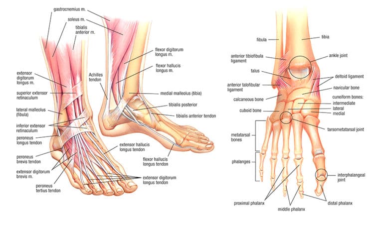
The hindfoot includes the heel (calcaneus) and the ankle bone (talus). The calcaneus acts as the primary point of contact between your foot and the ground. The Achilles tendon attaches to the back of the heel and connects your calf muscles to your foot, allowing you to push off the ground when walking or running.
The talus connects to both the tibia and fibula bones of your leg, forming the ankle joint, which allows the foot to move up and down. A complex network of ligaments supports the ankle and heel, ensuring stability while allowing movement.
Pain in the hindfoot often results from conditions like Achilles tendinitis, heel spurs, or ankle sprains. If you experience persistent pain in these areas, your podiatrist can provide treatments ranging from physical therapy to surgical intervention.
Muscles and Tendons of the Foot
More than 30 muscles in your foot control movement, stability, and balance. These muscles work with tendons, which connect muscle to bone, to move your toes and ankles. There are two main types of muscles in the foot: intrinsic muscles (inside the foot) and extrinsic muscles (located in the lower leg but affecting foot movement).
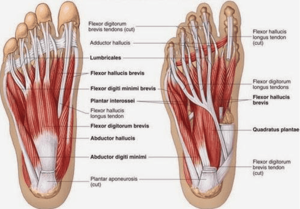
1. Intrinsic Muscles
The intrinsic muscles are small but crucial for foot function. They stabilise the arch, move the toes, and help control balance when walking or standing. Common injuries to these muscles include overuse and strain, which can cause pain and swelling.
2. Extrinsic Muscles
Extrinsic muscles start in the lower leg and control the foot through long tendons. These muscles allow you to lift, push, and rotate your foot. Some key tendons include:
- The Achilles tendon: The largest tendon in your body, it plays a vital role in walking, running, and jumping. Achilles tendinitis, a common injury, occurs when the tendon becomes inflamed due to overuse.
- The tibialis posterior tendon: This tendon helps maintain the arch of the foot. Dysfunction in this tendon can lead to flat feet and pain.
- The peroneal tendons: These run along the outside of the ankle and stabilise the foot when walking or running on uneven surfaces.
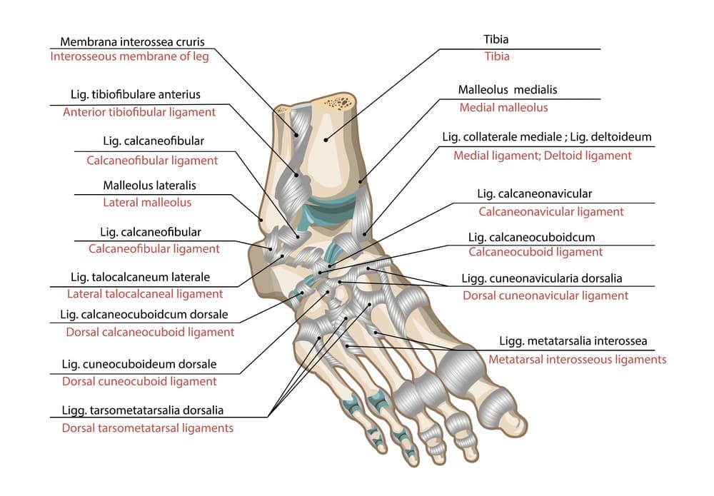
Ligaments in the Foot
Ligaments connect bones to other bones, providing stability and support. Several ligaments hold the bones of your foot together and ensure proper movement. Some of the key ligaments include:
- The plantar fascia: This ligament supports the arch and absorbs shock as you walk or run. Overstretching or overuse can cause plantar fasciitis, a painful condition often treated with stretches, orthotics, or other therapies.
- The deltoid ligament: Found on the inside of the ankle, it supports the ankle joint and prevents excessive movement. Sprains or tears in this ligament can cause significant ankle pain and instability.
- The lateral ligaments: These ligaments support the outside of the ankle and help prevent rolling. Ankle sprains often occur when these ligaments stretch or tear.
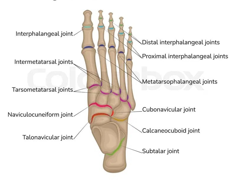
Joints of the Foot
More than 30 joints in your foot enable movement and flexibility. These joints play key roles in walking, running, jumping, and balancing.
1. Ankle Joint
The ankle joint (formed by the tibia, fibula, and talus bones) allows your foot to move up and down. This joint plays a major role in walking and running. If you injure your ankle, such as through sprains or fractures, you may experience swelling, pain, or instability.
2. Subtalar Joint
The subtalar joint sits below the ankle and allows side-to-side motion. This joint helps you maintain balance on uneven surfaces. Problems in this joint can cause foot pain and make walking difficult.
3. Metatarsophalangeal Joints
These joints connect the toes to the metatarsal bones. They play a crucial role in walking and running by allowing your toes to push off the ground. Arthritis, bunions, or other conditions can affect these joints, leading to pain and limited mobility.
Common Foot Conditions and How Foot Anatomy Contributes
Foot problems often arise due to an issue within the anatomy of the foot. For instance, a weak arch may cause plantar fasciitis, while tight calf muscles may contribute to Achilles tendinitis. Here are a few examples of how foot anatomy plays a role in common foot conditions:
- Plantar fasciitis: Tightness in the plantar fascia ligament causes pain in the heel and arch. Overuse, poor footwear, or flat feet may lead to this condition.
- Bunions: Misalignment of the metatarsophalangeal joint (where the big toe connects to the foot) can cause a bunion, a painful bony bump on the side of the foot.
- Achilles tendinitis: Inflammation in the Achilles tendon often stems from overuse, tight calf muscles, or improper footwear.
- Flat feet: Weak or dysfunctional arches lead to flat feet, which can cause pain in the feet, knees, hips, and back.
Caring for Your Feet
Understanding your foot’s anatomy can help you recognise problems early. Pay attention to any pain or discomfort in your feet, ankles, or toes. Proper footwear, regular stretching, and foot-strengthening exercises can help prevent many foot conditions. If you notice persistent pain, swelling, or deformities, a podiatrist can diagnose and treat the problem before it worsens.
At ModPod Podiatry, we provide expert care for all foot and ankle issues. We have Podiatrist’s in Sydney and can give you advice on:
Conclusion
Your foot plays a crucial role in supporting your body and enabling movement. Understanding the anatomy of the foot empowers you to take better care of it and seek help when needed. From the bones and muscles to the ligaments and joints, every part of your foot works together to keep you moving.
If you experience foot pain or discomfort, don’t wait. Visit ModPod Podiatry for professional care tailored to your needs. We’re here to help you maintain healthy, pain-free feet.